High Ankle Sprain

Do you know that high ankle sprain has forced many sportsmen and women out of their loved profession? Yet a lot more are ending their careers prematurely as a result of this monster. It is disheartening to see your loved football idol or a lawn tennis star suddenly take the exit just because of an injury everyone had considered to be a minor one. Well, that is the reality we all have to put up with. High ankle sprain is really a threat to everyone – sportsmen and the laymen alike. High ankle sprain comes from various causes; it could be sports related, a result of placing one’s step wrongly, skidding, a poor lifting technique – just anything.
What is High Ankle Sprain?
High ankle sprain is the sprain of the syndesmotic ligaments that link the tibia and fibula in the leg, which eventually lead to a mortise and tenon joint for the ankle.
Like the name suggests, high ankle sprains are situated above the ankle, and they make up about fifteen percent of all ankle sprains. While the lateral ankle sprain occurs as a result of an inward twisting of the ankle, the high ankle sprain is caused by the external rotation of the lower leg and foot.
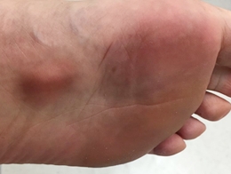
Symptoms
The following symptoms are common with high ankle sprain:
- Severe pain
- Minor swelling
- Inability to walk
- Pain on attempt to rotate the ankle outward
- Significant bruising
- Muscle spasm
- Cold foot or paresthesia (as a result of neurovascular involvement).
Causes of High Ankle Sprain
High ankle sprain has multiple causes from sports to road traffic accidents. In a nutshell, here are a couple causes of high ankle sprain that you need to familiarize yourself with.
- Hard tackles in a football game
- Wrong stepping
- Collision sports like wrestling, American football, Lacrosse, rugby, ice hockey, etc.
- Skidding
- Road traffic accident
- Fall from a height
- High velocity trauma
- High-heeled shoes
Diagnosis
The diagnosis of high ankle sprain is made from the presenting signs and symptoms of the injury as well as after a few medical investigations. In some cases, it is possible to arrive at a diagnosis without the need for further investigations, especially if the physician is highly experienced. However, for the sake of clarity, here are a few steps to take toward arriving at the right diagnosis of high ankle sprain.
Signs and symptoms
The presenting signs and symptoms would be enough in some cases. This is because the classical symptoms are a little different from those of fracture. Some of the signs to look out for include pain in the outside-front of the ankle (usually above the ankle), high discomfort when external rotation is applied to the leg, calf pain, swelling in a few cases, tenderness, bruising, difficulty walking, burning sensation on the affected site, etc. However, all of the aforementioned signs are also there in a fracture. But some of the signs and symptoms of fracture that are absent in high ankle sprain include crepitation, shortening of the affected limb, disfigurement of limb, swelling (in some cases), and a few others.
The squeeze test
This involves compressing the tibia and the fibula midpoint above the calf. The compression will certainly produce pains, thereby giving an idea of the type of injury. However, this alone may not be sufficient to arrive at a conclusive diagnosis. Other tests have to be performed before an accurate diagnosis is made.
Dorsiflexion with compression
When the examiner or doctor compresses the internal and external malleolus, one sign to look out for is whether the injured person would dorsiflex the foot. Should this happen, it is a sign that what the person has could possibly be a high ankle sprain.
X-ray
An x-ray of the area will reveal a widening of the tibia and fibula mortise, or a fracture of the medial malleolus. A Maisonneuve fracture will show a potentially unstable or a stable injury. However, normal x-rays hardly show significant ligament injury.
Ultrasound
An ultrasound of the leg will reveal a significant injury to the ligament that supports the tibia and fibula.
MRI
Magnetic Resonance Imaging (MRI) also gives a very clear picture of the level of injury or tear that has occurred to the distal tibiofibular ligaments. With this or the ultrasound, diagnosis can be made without many doubts.
Differential diagnosis
Differential diagnosis has to be made to rule out certain conditions. Usually, the differential diagnosis of high ankle sprain includes deltoid ligament sprain, Weber fractures, lateral ankle sprain, and Maisonneuve fracture.
Syndesmotic sprains can sometimes be accompanied by various other injuries, including osteochondral lesion of the distal tibiofibular joint, bone bruises, etc.
Treatment
The treatment of high ankle sprain is based on the severity of the injury. Healing time can be as short as a few days to as long as up to six months. All ligaments injuries require early intervention and quick control of swelling. The use of the “RICE” technique has proven to be a better way to handle the problem in the early stage.
Rest
In deciding whether to rest the leg, certain factors need to be considered first – if the ankle is stable or unstable. This can be answered based on the result of the imaging tests as well as clinical assessment of the situation.
The second factor to consider is the level of weight-bearing that is allowed. This also depends on the level of stability. If imaging findings and patient’s comfort when weight is applied to the leg are okay, then there will be no alarm. But in a situation where imaging suggests severe injury or there is pain and discomfort when weight is applied to the leg, there would be a need to rest the leg in order to recover.
Ice
Applying ice packs to the affected part immediately after the injury helps to minimize tissue inflammatory reaction and consequently leads to a reduction in the tendency for the leg to swell. Besides, the ice also helps in reducing pain experienced with the injury.
Compression
This involves wrapping, application of splint or cast. The idea behind this is to create immobilization of the affected area and allow it some time to heal itself. Immobilization in the form of short leg cast application is also important especially if there is a broken fibula, in order to allow it enough healing time.
Elevation
Elevation of the leg is equally necessary to improve venous blood return and reduce swelling. Elevating the leg also helps reduce platelet aggregation, thereby reducing swelling also.
Anti-inflammatory medication
Anti-inflammatory medications (such as Ibuprofen, Aspirin, and Diclofenac) and natural creams like Arnica can help to significantly minimise pain and swelling. You should, however, avoid anti-inflammatory medications in the first 48 to 72 hours of the injury in order to reduce the possibility of increased bleeding.
Surgery
Surgery may be indicated when there is marked instability as a result of a disruption of the normal tibiofibular relationships. If radiography reveals the presence of a fracture, surgery may also be needed. Other conditions that may require surgical intervention include scarring within the syndesmosis, latent instability, calcification in the syndesmosis, etc.
Recovery
Recovery from high ankle sprain takes a few days to as much as six months. Basically, recovery depends on the extent of the ligament damage. For full recovery to be achieved there may be need for resting of the leg, ice application, compression and elevation, as earlier discussed. Besides, physiotherapy is required to enable the physiotherapist to take the person through some physical therapy exercises.
Physiotherapy
The injured person may require the services of a physiotherapist in the long run. He has to be subjected to some physical therapy in order to prevent some complications such as chronic foot pain, or when he can’t walk after the primary injury seems to have healed. Other things that could be done include the use of custom orthoses, or walking boots. Crutches may be needed to help reduce the weight-bearing effect on the injured part.
Early resistance exercise limits the level of muscle atrophy and weakness. Different types of exercises may be included in the recovery therapy – ankle weights, elastic bands, heel raise exercises, range of motion exercise, etc may be used in addition to a calf stretch. Another way to minimize muscle atrophy and weakness in the early stage is to make use of electrical stimulation and isometric strengthening.
When to see a doctor
The presence of any of the signs and symptoms we talked about earlier should move you to see a doctor immediately in order to ascertain the extent of the injury. If you experience a burning sensation, a lump over the affected area, knee pain, or you can’t walk, then you need to see the doctor without wasting any time. You should never stay at home until the injury becomes complicated.
Conclusion
High ankle sprain is commoner among people involved in body contact sports; it accounts for 15% of all forms of ankle sprains. It can cause long term complications if not properly handled by a professional. However, with early intervention, it can be treated effetively and the injured person returns to his normal activities.